I. Causes of Retinal Detachment in Dogs
1.Genetic Factors
Some dog breeds have a genetic predisposition to congenital retinal dysplasia. Small breeds such as Pomeranians, Chihuahuas, and Shih Tzus may have inherent defects in the structure and function of their retinas, making the retinas prone to detachment. Retinal problems caused by genetic factors may gradually manifest in the dog's puppyhood.
2.Traumatic Factors
Physical Impact: When dogs are playing, running, or fighting, their eyes may be subject to external force impacts, such as being hit by other objects or colliding with their companions. These impacts can make the retina be shaken or torn, thereby triggering retinal detachment.
Accidents: Accidents such as car crashes and falls from heights can cause severe trauma to a dog's eyes. Besides directly damaging the retina, they may also lead to pathological changes in other structures within the eyeball, further increasing the risk of retinal detachment.
2.Eyes Diseases
Uveitis: This is a common inflammatory eye disease. Inflammation can lead to changes in the composition and pressure of the intraocular fluid, affecting the normal attachment of the retina. Long-term uveitis may cause the connection between the retina and the eyeball wall to become loose, ultimately leading to retinal detachment.
Glaucoma: Elevated intraocular pressure is the main feature of glaucoma. High eye pressure can compress the optic nerve and retina, obstructing the blood circulation of the retina, which in turn leads to damage to the retinal nerve fiber layer, insufficient nutrition supply to the retinal tissue, and gradual atrophy and detachment.
Cataract: As cataract aggravates, the lens gradually becomes cloudy and swollen, exerting mechanical pressure on surrounding tissues, pushing the iris forward, narrowing the angle of the anterior chamber, leading to increased intraocular pressure. It also affects the normal metabolism of the vitreous body, promoting vitreous liquefaction and degeneration, increasing traction on the retina, and thus triggering retinal detachment.
Retinal Vascular Diseases: Conditions such as retinal vascular occlusion and diabetic retinopathy affect the blood supply to the retina, leading to retinal ischemia and hypoxia, which in turn cause degeneration and necrosis of retinal tissue, reducing the stability of the retina and making it more prone to detachment.
3.Systemic Disease Factors
Diabetes: Chronic high blood sugar can damage blood vessels throughout the body, including those in the retina, leading to retinal microvascular changes. These changes include hemorrhages, exudates, and the formation of new blood vessels, which can disrupt the normal structure and function of the retina, increasing the likelihood of retinal detachment.
Hypertension: High blood pressure can increase the pressure in the retinal arteries, putting greater stress on the vessel walls, making them more susceptible to conditions such as arteriosclerosis and spasms. This can affect the blood perfusion of the retina, leading to ischemia and hypoxia, which in turn can cause retinal changes. In severe cases, this can lead to retinal detachment.
II. Consequences of Retinal Detachment in Dogs

1.Decreased Vision
Progressive Blindness: Once retinal detachment occurs, a dog's vision will gradually deteriorate until it becomes blind. In the early stages of the disease, the dog may only have blurred vision for distant or close objects, but as the condition progresses, vision will become increasingly poor, eventually leading to a complete loss of visual perception.
Limited Mobility: The loss of vision greatly restricts a dog's mobility. They struggle to discern obstacles in their environment, often colliding with furniture, walls, and other objects while walking. Navigating stairs also becomes difficult and hazardous.
2.Visual Field Defects
Peripheral Vision Loss: Retinal detachment can cause a dog's field of vision to gradually narrow, starting from the periphery and moving towards the center. Dogs will progressively lose the ability to perceive objects and movements from the sides or behind them.
Hemianopia: Partial retinal detachment may also result in hemianopia in dogs, which means they can only see a part of their visual field, while the other half is completely blind.
3.Ocular Discomfort and Pain
Inflammatory Response: Retinal detachment is often accompanied by symptoms such as redness, pain, and tearing of the eye. Inflammation can cause dogs to feel ocular discomfort, leading to frequent blinking and rubbing of the eyes, which may even result in complications like corneal ulcers, further exacerbating ocular damage and pain.
Intraocular Pressure Changes: Retinal detachment can cause changes in intraocular pressure, and both high and low pressure can cause distress to dogs. High intraocular pressure can lead to a feeling of fullness and pain in the eye, causing compressive damage to the optic nerve; low intraocular pressure may lead to eyeball atrophy, affecting the normal shape and function of the eye.
III. How to Prevent Retinal Detachment in Dogs and Supplement

1.Regular Eye Examinations
Establish a Routine: Take your dog to a professional veterinary hospital for comprehensive eye examinations regularly. It is recommended to do this at least 1-2 times per year.
Increase Examination Frequency During Special Periods: For senior dogs, or those with systemic diseases such as diabetes or hypertension, eye examinations should be conducted every six months or quarterly. For dogs with a genetic history of eye diseases, it is important to closely monitor their eye health from a young age, with examinations every 3-4 months.
2.Supplement Recommendations
While there are no specific supplements that can guarantee the prevention of retinal detachment, certain nutrients can support overall eye health. These include:
Antioxidants: Vitamins C and E can help protect the retina from damage caused by free radicals.
Omega-3 Fatty Acids: These can support the health of the retina and overall eye function.
Lutein and Zeaxanthin: These carotenoids are concentrated in the retina and can help protect it from light-induced damage.
Taurine: An amino acid that is essential for the health of the retina, especially in cats, but can also be beneficial for dogs.
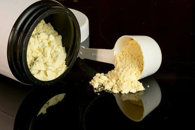
Best supplements recommended for dogs' health . For more information, please click the pictures.
![]()

Noora 8 in 1 contains vitamin E and vitamin C, and Selenium, good for vision health for you pets. .
Advantages: Specifically designed for dogs, this supplement provides comprehensive health support, including for joints, muscles, cardiovascular health, skin, gut, vision health and immune system.
Appearance/Taste: This product is available in chewable tablets that have a chicken liver taste to make it more appealing for dogs.
Size: 2.5 grams each tablet, 50 tablets in each bottle, a total of 125g (4.41 )

Noora Daily Multi contains vitamin A vitamin C and vitamin E, good for vision health
Advantages: Noora Daily Multi is a comprehensive health supplement designed to complement today's dog diets by providing 21 essential nutrients. It supports overall health maintenance and well-being in dogs of all ages, ensuring they receive a balanced intake of vital vitamins and minerals.
Appearance/Taste: The supplement comes in a vegetable flavor, which is appealing to dogs and encourages consumption.
Size: Each chew is formulated to be 3.5 grams, making it a manageable size for dogs to chew comfortably.
Quantity: 60 chews in each bottle, a total of 210 g (7.41 oz).
3.Active Prevention and Treatment of Eye Diseases
Prompt Inflammation Treatment: If you notice that your dog has eye inflammation, such as conjunctivitis, keratitis, or uveitis, it is important to take them to the hospital for treatment immediately. Follow the veterinarian's advice on using medications, administer eye drops, and give oral medications as scheduled to ensure that the inflammation is completely controlled. This prevents the inflammation from spreading to the retina and causing retinal detachment.
Control Intraocular Pressure: Regularly measure your dog's intraocular pressure, especially for those with glaucoma or a family history of glaucoma. Closely monitor changes in intraocular pressure. If an increase in pressure is detected, immediate measures should be taken to reduce it, such as using intraocular pressure-lowering medications or undergoing surgical treatment, to prevent damage to the optic nerve and retina from high pressure.
Monitor Cataract Progression: For dogs with cataracts, regular follow-ups are necessary to observe the progression of the condition. In the early stages of cataracts, the progression can be delayed with medication. When cataracts significantly impair vision and meet the criteria for surgery, timely cataract surgery should be performed to prevent retinal detachment due to changes in the internal eye structure caused by cataracts.